
Nature 2022 Oct;610(7931):319-326. doi: 10.1038/s41586-022-05277-w. Epub 2022 Oct 12.
Maturation and circuit integration of transplanted human cortical organoids (移植されたヒト皮質オルガノイドの成熟と回路統合)
Omer Revah # 1 2, Felicity Gore # 1 3, Kevin W Kelley # 1 2, Jimena Andersen 1 2, Noriaki Sakai 1, Xiaoyu Chen 1 2, Min-Yin Li 1 2, Fikri Birey 1 2, Xiao Yang 1 2 4, Nay L Saw 5, Samuel W Baker 6, Neal D Amin 1 2, Shravanti Kulkarni 1 2, Rachana Mudipalli 1 3, Bianxiao Cui 4, Seiji Nishino 1, Gerald A Grant 7, Juliet K Knowles 8, Mehrdad Shamloo 5 7, John R Huguenard 8, Karl Deisseroth 1 3 9, Sergiu P Pașca 10 11
Sergiu P. Pașca(セルジュ・P・パスカ 1982年1月30日生まれ)は、ルーマニア系アメリカ人の科学者であり、カリフォルニア州のスタンフォード大学の医師である。神経科学者、幹細胞生物学者であり、現在はニューヨーク幹細胞財団のロバートソン研究員である。彼はスタンフォード神経科学研究所、スタンフォードバイオXの一部であり、スタンフォードのChEM-H研究所のフェローである。Pașcaは、医学と科学におけるニューヨークタイムズのビジョナリーにリストされており、彼はVlicek Foundationから2018 Vilcek Award for Creative Biomedical Promiseを授与されている。 2022年に彼は実験室で人間の脳をリバースエンジニアすることについてTEDトークをしている。 (Wikipediaより)
Affiliations
1Department of Psychiatry and Behavioral Sciences, Stanford University, Stanford, CA, USA.
2Stanford Brain Organogenesis, Wu Tsai Neurosciences Institute and Bio-X, Stanford University, Stanford, CA, USA.
3Department of Bioengineering, Stanford University, Stanford, CA, USA.
4Department of Chemistry, Stanford University, Stanford, CA, USA.
5Stanford Behavioral and Functional Neuroscience Laboratory, Wu Tsai Neurosciences Institute, Stanford University, Stanford, CA, USA.
6Department of Comparative Medicine, Stanford University, Stanford, CA, USA.
7Department of Neurosurgery, Stanford University, Stanford, CA, USA.
8Department of Neurology and Neurological Sciences, Stanford, CA, USA.
9Howard Hughes Medical Institute, Stanford University, Stanford, CA, USA.
10Department of Psychiatry and Behavioral Sciences, Stanford University, Stanford, CA, USA. spasca@stanford.edu.
11Stanford Brain Organogenesis, Wu Tsai Neurosciences Institute and Bio-X, Stanford University, Stanford, CA, USA. spasca@stanford.edu. # Contributed equally.
神経発達:成熟して回路に統合される移植オルガノイド
今回S Pașcaらは、ラットの皮質に移植したヒト由来の皮質オルガノイドが、ロバストに成熟して宿主回路へと組み込まれ、行動に影響を与えるようになることを示している。
Abstract
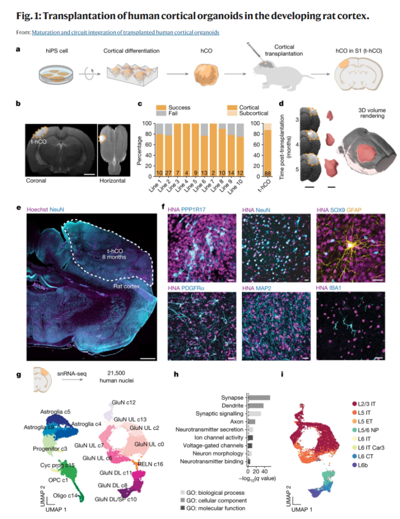
Self-organizing neural organoids represent a promising in vitro platform with which to model human development and disease1,2,3,4,5. However, organoids lack the connectivity that exists in vivo, which limits maturation and makes integration with other circuits that control behaviour impossible. Here we show that human stem cell-derived cortical organoids transplanted into the somatosensory cortex of newborn athymic rats develop mature cell types that integrate into sensory and motivation-related circuits. MRI reveals post-transplantation organoid growth across multiple stem cell lines and animals, whereas single-nucleus profiling shows progression of corticogenesis and the emergence of activity-dependent transcriptional programs. Indeed, transplanted cortical neurons display more complex morphological, synaptic and intrinsic membrane properties than their in vitro counterparts, which enables the discovery of defects in neurons derived from individuals with Timothy syndrome. Anatomical and functional tracings show that transplanted organoids receive thalamocortical and corticocortical inputs, and in vivo recordings of neural activity demonstrate that these inputs can produce sensory responses in human cells. Finally, cortical organoids extend axons throughout the rat brain and their optogenetic activation can drive reward-seeking behaviour. Thus, transplanted human cortical neurons mature and engage host circuits that control behaviour. We anticipate that this approach will be useful for detecting circuit-level phenotypes in patient-derived cells that cannot otherwise be uncovered.
この論文の要旨
自己組織化する神経オルガノイドは、ヒトの発生や疾患のモデルとなり得る有望なin vitroプラットフォームである。しかし、オルガノイドはin vivoに存在するような神経結合を欠くため、成熟に限界があり、行動を制御する他の回路との統合は不可能である。今回我々は、ヒト幹細胞由来の皮質オルガノイドを、新生の無胸腺ラットの体性感覚皮質に移植すると、それらが成熟した細胞タイプにまで発達し、感覚および動機に関連した神経回路に組み込まれることを示す。MRIによって、移植後のオルガノイドの成長が複数の幹細胞系列や動物にわたって見られることが分かり、単一核プロファイリングによって、皮質形成の進行と活動依存的な転写プログラムの出現が示された。実際、移植された皮質ニューロンはin vitroのものと比べて、より複雑な形態的、シナプス的、本質的な膜特性を示し、これにより、ティモシー症候群患者由来のニューロンで異状の発見が可能になった。解剖的・機能的な追跡によって、移植されたオルガノイドが、視床–皮質入力および皮質–皮質入力を受けることが明らかになり、神経活動のin vivo記録によって、これらの入力がヒト細胞で感覚応答を起こすことが実証された。さらに、皮質オルガノイドはラットの脳領域全体に軸索を伸ばしており、これらを光遺伝学に活性化すると報酬探索行動が誘発された。このように、移植ヒト皮質ニューロンは、成熟して、行動を制御する宿主動物の回路に関わるようになる。我々は、今回の手法が、他の方法では解明できない、患者由来の細胞の回路レベルでの表現型の検出に有用になると期待する。(Nature Japaより)
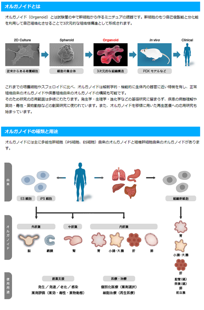
〇オルガノイドとは
〇解説
ラットに移植したヒトの脳のような小型構造体は、信号を送ったり、ラットのひげで拾った環境の手がかりに反応したりすることができることが、本研究により明らかになった。ヒトの幹細胞から育てたニューロンが、生きたネズミの神経細胞と相互作用できることを実証したこの研究は、ヒトの脳疾患の治療法を検証する方法につながる可能性がある。
ハイブリッドブレイン:ヒトの神経細胞を動物に移植することの倫理性
科学者たちは、脳オルガノイド(ヒト幹細胞から育てた脳のような小さな構造体)を使って、人間が発症する神経変性疾患や神経精神疾患を研究したいと考えている。しかし、オルガノイドが人間の脳を模倣するのはここまでである。血管が発達していないので、栄養を受け取ることができず、長くは生きられない。人間の乳児の脳では、神経細胞の成長や他の神経細胞との結合の発達は、感覚からの入力に基づくところがある。
最初のサルとヒトの胚がハイブリッド動物についての議論を再燃させる
倫理的な課題もある。齧歯類と人間のハイブリッドを作ると動物に害を与えるのではないか、あるいは人間のような脳を持った動物が生まれるのではないか、という懸念である。昨年、米国科学・工学・医学アカデミーが組織した委員会は、ヒトの脳オルガノイドはまだ原始的であり、意識を持ち、人間のような知能を持ち、その他の法的規制を必要とするような能力を身につけることはできないと結論づけた報告書を発表している。パスカによれば、彼のチームのオルガノイド移植は、ラットに発作や記憶障害などの問題を起こさず、動物の行動にも大きな変化はないようだという。
しかし、全米アカデミーのパネルのメンバーであるアルロッタは、科学の進歩に伴って問題が発生する可能性があると言う。「一度議論して、そのままにしておくわけにはいかないのです』と彼女は言う。また、ヒトオルガノイドに関する懸念は、神経疾患や精神疾患を持つ人々のニーズと天秤にかける必要があるとも述べている。脳内オルガノイドやヒトと動物のハイブリッド脳は、これらの疾患の根底にあるメカニズムを明らかにし、研究者が統合失調症や双極性障害などの治療法をテストできるようにする可能性がある。「私たちは社会人として、できる限りのことをする責任があると思います」と、アルロッタは言う。
“Journal Club (December 12, 2022)” への1件のフィードバック