SCIENCE 23 Jun 2022 Vol 377, Issue 6602 DOI: 10.1126/science.abo0924
Connectomic comparison of mouse and human cortex. (マウスとヒトの大脳皮質のコネクトーム比較)
Loomba S, Straehle J, Gangadharan V, Heike N, Khalifa A, Motta A, Ju N, Sievers M, Gempt J, Meyer HS, Helmstaedter M.
Department of Connectomics, Max Planck Institute for Brain Research, Frankfurt, Germany. (マックスプランク脳研究所 コネクトミクス研究部門)
Summary-ヒトとマウスの違い-
過去数十年の間に、マウスは脳研究のモデル生物となった。イオンチャネルやシナプス受容体などの脳の主要な分子構成がヒトと進化的に類似していることから、大脳皮質神経回路についても同様の類似性が想定されてきた。しかし、回路構造が種間でどの程度、保存あるいは進化してきたかを明らかにするためには、シナプスレベルの高解像度のコネクトミクスの比較研究が必要である。Loombaらは、三次元電子顕微鏡(シリアルブロックフェイス走査電子顕微鏡法(SBEM、SBSEM、およびSBFSEM*))を用いて、マウスとヒト/マカクサル大脳皮質のシナプス結合性を比較した。ヒトの細胞はマウスの神経細胞に比べてはるかに大きく、数も多いが、平均してより多くのシナプスを受け取ってはいない。また、ヒトの大脳皮質にはマウスの3倍もの介在ニューロンがあるにもかかわらず、興奮と抑制の比率は両種間でほぼ同じであることがわかった。
*シリアルブロックフェイス走査電子顕微鏡法(SBEM、SBSEM、およびSBFSEM)
Abstract
The human cerebral cortex houses 1000 times more neurons than that of the cerebral cortex of a mouse, but the possible differences in synaptic circuits between these species are still poorly understood. We used three-dimensional electron microscopy of mouse, macaque, and human cortical samples to study their cell type composition and synaptic circuit architecture. The 2.5-fold increase in interneurons in humans compared with mice was compensated by a change in axonal connection probabilities and therefore did not yield a commensurate increase in inhibitory-versus-excitatory synaptic input balance on human pyramidal cells. Rather, increased inhibition created an expanded interneuron-to-interneuron network, driven by an expansion of interneuron-targeting interneuron types and an increase in their synaptic selectivity for interneuron innervation. These constitute key neuronal network alterations in the human cortex.
ヒト大脳皮質にはマウス大脳皮質の1,000倍もの神経細胞が存在するが、これらの種間のシナプス回路の違いについては未だ十分に理解されていない。本研究では、マウス・マカク・ヒトの大脳皮質のサンプルを三次元電子顕微鏡で観察し、その細胞型構成とシナプス回路構造を調べた。マウスに較べてヒトでは抑制性ニューロン(IN)が2.5倍増加していたが、軸索の接続確率も変化していたことから、ヒト錐体細胞への抑制性-興奮性シナプス入力バランスは相応に増加しなかった。増加したINの主な細胞腫は双極性抑制性ニューロン(BP IN)であり、BP INがIN-IN間の回路を拡大させていたことが明らかになった。これらの回路の発達は、大脳皮質の進化において重要であり、今後更なる研究の対象となることが予想される。
〇研究成果
ヒトの側頭・前頭皮質、マカクの側頭・頭頂皮質から3次元電子顕微鏡データを取得した。これらのデータから、コネクトーム再構成を行い、マウス大脳皮質の5つのコネクトームと比較した。これらのデータをもとに、マカクルとヒトの大脳皮質における介在ニューロン集団が、マウスのそれと比べて約2.5倍に拡大していることの影響を明らかにすることができた。予想に反して、マカクルとヒトの大脳皮質では、錐体細胞の抑制性と興奮性のシナプスバランスは大きく変化しなかった。むしろ、介在ニューロン集団は、他の介在ニューロンの神経支配に選好性をもつ双極型介在ニューロンが選択的に拡大していた。マウスからヒトにかけて介在ニューロン神経支配への選好性がさらに増加していた。これらの変化はそれぞれ数倍であり、マウスにはまばらにしか存在しない介在ニューロン間ネットワークが、ヒト大脳皮質では実質的には約10倍に拡大したことになる。しかし、錐体細胞へのシナプス入力の総量は、大脳皮質の3倍厚くなっても変化せず、むしろ、マウスでは約12,000のシナプス入力が、ヒトでは約15,000に緩やかに増加することが分かった。
〇結論
大脳皮質の主要細胞である錐体細胞は、1億年の進化的分岐を経ても、抑制性入力と興奮性入力のバランスと総シナプス入力はほぼ一定であり、特に神経細胞ネットワークサイズが1000倍に拡大し、抑制性介在ニューロンがマウスからヒトへ2.5倍増加したことが注目される。むしろ、マウスからヒトへの重要なネットワークの変化は、マウスにはほとんど存在しないが、ヒトの皮質ネットワークのかなりの部分を構成する介在ニューロン間のネットワークが、ほぼ1桁拡大したことである。この新しいネットワークが、主に既存の神経細胞の種類の拡大によって作られたのか、それとも新しい介在ニューロンのサブタイプの作成に関係しているのか、さらなる研究が必要である。ヒトの大脳皮質でこのネットワークが発見されたことで、正常脳や疾病におけるその機能の詳細な分析が可能になった。
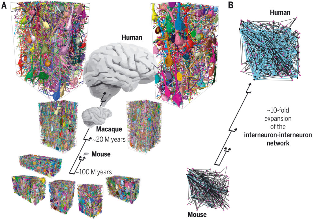
(A) Automated reconstructions of all neurons with their cell bodies in the volume shown, using random colors. The analyzed connectomes comprised a total of ~1.6 million synapses. Arrows indicate evolutionary divergence: the last common ancestor between human and mouse, approximately 100 million years ago, and the last common ancestor between human and macaque, about 20 million years ago. (B) Illustration of the about 10-fold expansion of the interneuron-to-interneuron network from mouse to human.
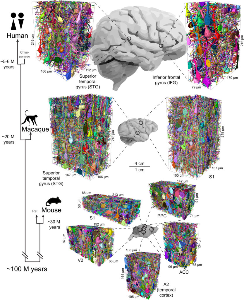
Dense connectomic reconstructions from L2/3 of five cortical areas of mouse (bottom, n = 5 individuals) and from four cortical areas of macaque and human (n = 3 individuals). There are matched cortical areas (A2 and STG) across all three species and paired samples from S1 (mouse and macaque). A total of 202,954 axons and 1,618,129 synapses were analyzed (Materials and methods). The raw 3D EM data of mouse datasets S1, V2, PPC, and ACC were previously published (55), but their dense reconstruction have not. (Left) Simplified phylogenetic tree [based on (100)] indicating time to last common ancestor between human (Homo sapiens), rhesus macaque (Macaca mulatta), and mouse (Mus musculus). Scale bars apply to the brain sketches. S1, primary somatosensory cortex; A2, secondary auditory cortex; V2, secondary visual cortex; PPC, posterior parietal cortex; ACC, anterior cingulate cortex; STG, superior temporal gyrus; IFG, inferior frontal gyrus.
論文紹介:Y.W. (医学類4年)