Nature Article Published: 01 October 2021 volume 598, pages483–488 (2021)
Regulation of prefrontal patterning and connectivity by retinoic acid (レチノイン酸による前頭前野の形態形成と神経回路の制御)
Mikihito Shibata, Kartik Pattabiraman, Belen Lorente-Galdos, David Andrijevic, Suel-Kee Kim, Navjot Kaur, Sydney K. Muchnik, Xiaojun Xing, Gabriel Santpere, Andre M. M. Sousa & Nenad Sestan*
- Department of Neuroscience, Yale School of Medicine, New Haven, CT, USA
- Department of Genetics, Yale School of Medicine, New Haven, CT, USA
- Yale Genome Editing Center, Yale School of Medicine, New Haven, CT, USA
- Department of Comparative Medicine, Yale School of Medicine, New Haven, CT, USA
- Department of Psychiatry, Yale School of Medicine, New Haven, CT, USA
- Program in Cellular Neuroscience, Neurodegeneration and Repair, Yale School of Medicine, New Haven, CT, USA
- Kavli Institute for Neuroscience, Yale University, New Haven, CT, USA
*Nenad Sestanは、Yale大学医学部の教授である。
Abstract
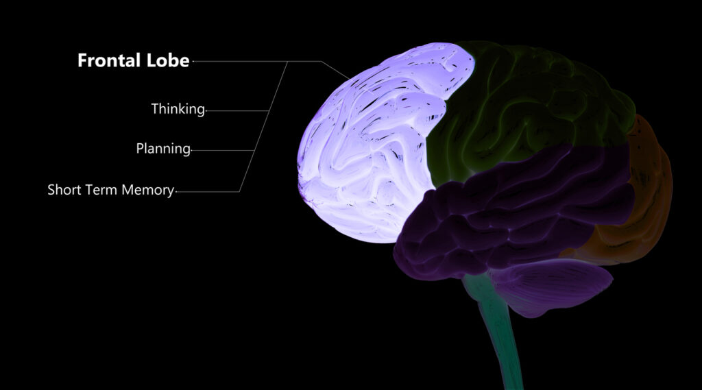
The prefrontal cortex (PFC) and its connections with the mediodorsal thalamus are crucial for cognitive flexibility and working memory1 and are thought to be altered in disorders such as autism2,3 and schizophrenia4,5. Although developmental mechanisms that govern the regional patterning of the cerebral cortex have been characterized in rodents6,7,8,9, the mechanisms that underlie the development of PFC–mediodorsal thalamus connectivity and the lateral expansion of the PFC with a distinct granular layer 4 in primates10,11 remain unknown.
Here we report an anterior (frontal) to posterior (temporal), PFC-enriched gradient of retinoic acid, a signalling molecule that regulates neural development and function12,13,14,15, and we identify genes that are regulated by retinoic acid in the neocortex of humans and macaques at the early and middle stages of fetal development. We observed several potential sources of retinoic acid, including the expression and cortical expansion of retinoic-acid-synthesizing enzymes specifically in primates as compared to mice.
Furthermore, retinoic acid signalling is largely confined to the prospective PFC by CYP26B1, a retinoic-acid-catabolizing enzyme, which is upregulated in the prospective motor cortex. Genetic deletions in mice revealed that retinoic acid signalling through the retinoic acid receptors RXRG and RARB, as well as CYP26B1-dependent catabolism, are involved in proper molecular patterning of prefrontal and motor areas, development of PFC–mediodorsal thalamus connectivity, intra-PFC dendritic spinogenesis and expression of the layer 4 marker RORB. Together, these findings show that retinoic acid signalling has a critical role in the development of the PFC and, potentially, in its evolutionary expansion.
要約
前頭前皮質(PFC)とその視床背内側核(MD)との結合は、認知の柔軟性や作業記憶に重要であり、自閉症や統合失調症などの疾患では変化していると考えられている。大脳皮質の領域パターニングを支配する発生メカニズムは、げっ歯類では明らかにされているが、霊長類ではPFCと視床背内側核(MD)の結合性の発生過程や、第4層(内顆粒層)を特徴とするPFCの外側への拡大がどのようにして行われるのかは不明である。
筆者たちは、神経の発達と機能を制御するシグナル分子であるレチノイン酸が、前側から側方にかけて勾配を伴って、PFCに豊富に分布していることを報告している。また胚発生初期から中期にかけて、ヒトやマカクザルの大脳皮質においてレチノイン酸により制御される遺伝子を同定した。さらにげっ歯類と比較して、霊長類で特異的にレチノイン酸合成酵素が発現し皮質が拡大していることなど、複数のレチノイン酸の潜在的供給源を見出した。
さらに、レチノイン酸のシグナルは、レチノイン酸の異化酵素であるCYP26B1が将来の運動野で発現することにより、PFCに限定されることがわかった。マウスの遺伝子ノックアウトにより、レチノイン酸受容体RXRGおよびRARBを介したレチノイン酸シグナルと、CYP26B1に依存した代謝作用が、前頭葉および運動野の適切な分子パターニング、PFC-視床下部間の連結性の発達、PFC内の樹状突起のスパイン形成、第4層マーカーRORBの発現に関与していることが明らかになった。これらの結果は、レチノイン酸シグナルがPFCの発達に重要な役割を果たしていることを示しており、PFCの進化にも重要な役割を果たしている可能性がある。
NEWS AND VIEWS
01 October 2021
The genetic symphony underlying evolution of the brain’s prefrontal cortex
The prefrontal cortex of the human brain is larger than that of other species. Comparisons of mouse, macaque and human brains uncover some of the genetic and molecular factors behind these differences.
Jenelle L. Wallace & Alex A. Pollen
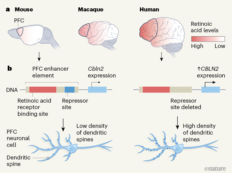
図1:マウス、マカク、ヒトの脳の大脳皮質の違い。マウス、マカク、ヒトの脳を比較した。その結果、レチノイン酸という分子の濃度は、3種とも脳の発生段階で大脳皮質の前部から後部に向かって勾配を示し、全体としてはヒトが最も高く、マウスが最も低いことがわかった。ヒトの前頭前野(PFC)は、マカクやマウスに比べて拡大しており、脳の別の構造体である視床との相互結合の数もマウスのPFCよりも多い(結合は図示されていない)。このエンハンサーには、レチノイン酸受容体やSOX5タンパク質が結合して、それぞれ遺伝子の発現を促進したり抑制したりする部位が含まれている。ヒトでは、抑制部位の一部が欠失しており、CBLN2の発現が増加している。マウスPFCの神経細胞は、シナプス結合を形成する樹状突起スパインと呼ばれる構造の密度が、ヒトのPFCの神経細胞に比べて低い。
“Journal Club (November 15, 2021)” への2件のフィードバック