Article Open Access Scientific Reports volume 11, Article number: 12350 (2021)
Published: 11 June 2021
Dendritic spines are lost in clusters in Alzheimer’s disease
Mite Mijalkov, Giovanni Volpe, Isabel Fernaud-Espinosa, Javier DeFelipe, Joana B. Pereira & Paula Merino-Serrais
- Laboratorio Cajal de Circuitos Corticales (CTB), Centro de Tecnología Biomédica, Universidad Politécnica de Madrid (マドリード工科大学), Campus Montegancedo S/N, Pozuelo de Alarcon, 28223, Madrid, Spain *マドリード工科大学は、スペイン・マドリードにある公立大学である。18世紀に開校した工学と建築学の技術学校が1971年に合併して開校した。
- Departamento de Neurobiología Funcional y de Sistemas, Instituto Cajal (カハール研究所), CSIC, Avenida Doctor Arce, 37, 28002, Madrid, Spain *カハール研究所は1932年に設置された、スペイン国立研究評議会に所属する神経生物学の研究センターである。
- Scientific Reportsは、Nature Research社によって刊行されているオープンアクセス学術雑誌である。自然科学の全分野を網羅しており、論文の重要性やインパクトではなく、科学的正当性のみを評価することを目的とする。
Abstract
Alzheimer’s disease (AD) is a progressive neurodegenerative disorder characterized by a deterioration of neuronal connectivity. The pathological accumulation of tau in neurons is one of the hallmarks of AD and has been connected to the loss of dendritic spines of pyramidal cells, which are the major targets of cortical excitatory synapses and key elements in memory storage. However, the detailed mechanisms underlying the loss of dendritic spines in individuals with AD are still unclear. Here, we used graph-theory approaches to compare the distribution of dendritic spines from neurons with and without tau pathology of AD individuals. We found that the presence of tau pathology determines the loss of dendritic spines in clusters, ruling out alternative models where spine loss occurs at random locations. Since memory storage has been associated with synaptic clusters, the present results provide a new insight into the mechanisms by which tau drives synaptic damage in AD, paving the way to memory deficits through alterations of spine organization.
アルツハイマー型認知症(AD)は、神経細胞の結合性の低下を特徴とする進行性の神経変性疾患である。アルツハイマー病では、神経細胞内にタウ(Tau 注1)が蓄積し、錐体細胞の樹状突起が消失することが知られている。しかし、AD患者における樹状突起の消失の詳細なメカニズムはまだ明らかになっていない。今回、筆者たちはグラフ理論(注2)を用いて、AD患者のタウ病理の有無にかかわらず、神経細胞の樹状突起の分布を比較した。その結果、タウ病理の存在が樹状突起スパイン(注3)の消失をクラスター化させることを発見し、スパインの消失がランダムに起こるという別のモデルを否定した。記憶の保存はシナプスのクラスターと関連していることから、今回の結果は、タウがADのシナプス障害を引き起こすメカニズムについての新たな知見を提供するものであり、スパインの組織化の変化による記憶障害への道を開くものである。
注1: アルツハイマー病患者の脳では、異常にリン酸化された神経軸索内のタウ(Tau)タンパク質が樹状突起スパインに侵入し、神経変性が引き起こされている。
注2: グラフ理論(Graph theory)は、ノード(節点・頂点)の集合とエッジ(枝・辺)の集合で構成されるグラフに関する数学の理論を指す。
注3: 樹状突起スパイン(樹状突起棘: Dendritic spine)とは、神経細胞の樹状突起から突き出ている小区画で、脳の興奮性シナプスの入力を受ける棘状の突起である。
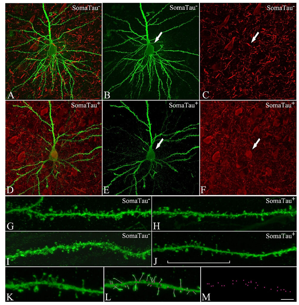
CA1 pyramidal neurons with and without tau pathology. Confocal microscopy pictures obtained after combining the channels acquired separately for LY (green) and phospho-tau AT8 (red), showing (A–F) neurons and (G–J) their basal dendrites, (A–C,G–H) with a soma free of phospho-tau AT8 (SomaTau-) and (D–F,I–J) with phospho-tau AT8 in an intermediate stage of neurofibrillary pathology (SomaTau +). The position of the soma is indicated with an arrow in (B,C) and (E,F). (K) High resolution image of a dendritic segment indicated with a bracket in J. (L) The same representative image as in (K) showing all the spines along the dendrite marked with a white line and pink dots for their insertion points. (M) 3D spatial distribution of all spines insertion points. Scale bar shown in (M) indicates 12 µm in (A–F), 5 µm in (G–J), and 3 µm in (K–M).
https://www.nature.com/articles/s41598-021-91726-x/figures/1