PNAS RESEARCH ARTICLE NEUROSCIENCE June 6, 2023 120 (24) e2220200120
Heterogeneous growth of the insula shapes the human brain (島皮質の不均一な成長がヒトの脳を形作る)
Arka N Mallela 1, Hansen Deng 1, Ali Gholipour 2 3, Simon K Warfield 2 3, Ezequiel Goldschmidt 2 4
- 1Department of Neurological Surgery, University of Pittsburgh Medical Center, Pittsburgh, PA 15213.
- 2Department of Radiology, Harvard Medical School, Boston, MA 02115.
- 3Department of Radiology, Boston Children’s Hospital, Boston, MA 02115.
- 4Department of Neurological Surgery, University of California San Francisco, San Francisco, CA 94143.
Significance
Buried in the depth of the Sylvian fissure, the insula not only has a flatter, less convoluted surface than the cerebral convexity, but also is the focal point around which the cerebrum is arranged. Existing models of cortical folding do not explain why the insula has a unique morphology, nor its central location in the cerebrum. Here, we quantitatively explore the unique morphology and development of the insula by analyzing in utero fetal and adult brain MRI and diffusion tractography. We show that the insula has both a divergent growth and folding trajectory than the other lobes. Specifically, the insula exhibits much lower surface area expansion, a main force driving cortical folding, compared to other lobes. A curved stream of radially migrating cells originates from the ventricular zone and curve around the lenticular nuclei to reach the insula. Differential expansion between the insula and other lobes results in the formation of the Sylvian fissure and thus defines the interlobar geometry of the human brain.
外側溝(シルヴィウス溝 )の奥深くに埋もれている島皮質は、大脳の凸部よりも平坦で凸凹の少ない表面を持つだけでなく、大脳が配置される中心点でもある。既存の大脳皮質褶曲モデルでは、島皮質が独特の形態を持つ理由や、大脳の中心に位置する理由は説明できない。ここでは、胎生期と成体期のMRIと拡散トラクトグラフィーを解析することで、島皮質の独特な形態と発達を定量的に探る。その結果、島皮質が他の脳葉と比較して、乖離した成長と折りたたみの軌跡を描くことがわかった。具体的には、島皮質は他の葉に比べて、皮質の褶曲を促進する主要な力である表面積の拡大が非常に小さい。放射状に移動する細胞の湾曲した流れは、脳室帯から発生し、レンズ状核の周りを曲がって島皮質に到達する。島皮質と他の葉との間の膨張の差はシルビウス裂の形成につながり、その結果、ヒトの脳の葉間幾何学が規定される。
Abstract
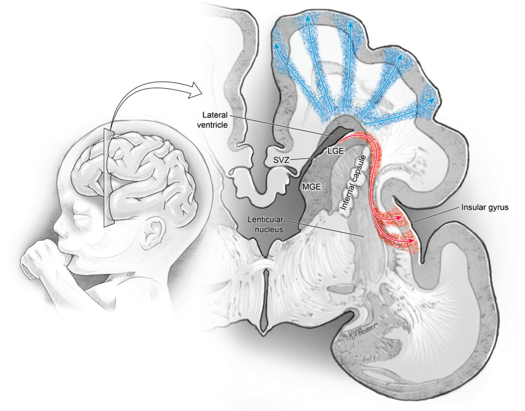
The human cerebrum consists of a precise and stereotyped arrangement of lobes, primary gyri, and connectivity that underlies human cognition [P. Rakic, Nat. Rev. Neurosci.10, 724-735 (2009)]. The development of this arrangement is less clear. Current models explain individual primary gyrification but largely do not account for the global configuration of the cerebral lobes [T. Tallinen, J. Y. Chung, J. S. Biggins, L. Mahadevan, Proc. Natl. Acad. Sci. U.S.A.111, 12667-12672 (2014) and D. C. Van Essen, Nature385, 313-318 (1997)]. The insula, buried in the depths of the Sylvian fissure, is unique in terms of gyral anatomy and size. Here, we quantitatively show that the insula has unique morphology and location in the cerebrum and that these key differences emerge during fetal development. Finally, we identify quantitative differences in developmental migration patterns to the insula that may underlie these differences. We calculated morphologic data in the insula and other lobes in adults (N = 107) and in an in utero fetal brain atlas (N = 81 healthy fetuses). In utero, the insula grows an order of magnitude slower than the other lobes and demonstrates shallower sulci, less curvature, and less surface complexity both in adults and progressively throughout fetal development. Spherical projection analysis demonstrates that the lenticular nuclei obstruct 60 to 70% of radial pathways from the ventricular zone (VZ) to the insula, forcing a curved migration to the insula in contrast to a direct radial pathway. Using fetal diffusion tractography, we identify radial glial fascicles that originate from the VZ and curve around the lenticular nuclei to form the insula. These results confirm existing models of radial migration to the cortex and illustrate findings that suggest differential insular and cerebral development, laying the groundwork to understand cerebral malformations and insular function and pathologies.
ヒトの大脳は、脳葉、一次脳回(大脳の隆起部分)、神経結合の正確かつ定型的な配置で構成されており、それがヒトの認知を支えている [P. Rakic, Nat. Rev. Neurosci.10, 724-735 (2009)]。この配列の発達はあまり明らかではない。現在のモデルでは、個々の一次脳回形成は説明できるが、大脳葉のグローバルな配置はほとんど説明できない[T. Tallinen, J. Y. Chung, J. S. Biggins, L. Mahadevan, Proc. Natl. Acad. Sci. U.S.A.111, 12667-12672 (2014) and D. C. Van Essen, Nature385, 313-318 (1997)〕。] 外側溝(シルヴィア溝)の深部に埋もれている島皮質(insula)は、その脳回の解剖学的構造と大きさの点でユニークである。ここでは、島皮質が大脳の中でユニークな形態と位置を持つこと、そしてこれらの重要な違いが胎児期の発生過程で現れることを定量的に示す。最後に、これらの違いの根底にある可能性のある、島皮質への発生的移動パターンにおける定量的な違いを明らかにする。我々は、成人(N = 107)と胎内胎児脳アトラス(N = 81健常胎児)の島皮質と他の葉の形態学的データを計算した。胎内では、島葉は他の葉よりも一桁遅く成長し、成体でも胎児の発育過程でも、より浅い溝、より少ない曲率、より少ない表面の複雑さを示す。球面投影解析により、レンズ核が脳室帯(VZ)から島への放射状経路の60~70%を閉塞し、放射状の直接経路とは対照的に島への湾曲した移動を余儀なくされることが示された。胎児期の拡散トラクトグラフィーを用いて、我々はVZから発生し、レンズ状核の周囲で湾曲して島を形成する放射状グリアを同定した。これらの結果は、大脳皮質への放射状の移動に関する既存のモデルを確認するとともに、島と大脳の発達の違いを示唆する所見を示しており、大脳奇形と島機能および病態を理解するための基礎を築くものである。
Keywords: brain development; brain folding; fetal MRI; gyrification; insula.
Discussion
脳発生の第2期と第3期には、大脳皮質は滑らかな終脳小胞から、小葉と保存された脳溝と脳回がはっきりとしたパターンを持つ、折り畳まれた表面へと拡大する。脳の折り畳みに関する既存の物理モデルは、個々の回や溝の形成を説明することができるが、島皮質に関する脳の全体的な形状の形成は依然として説明されていない。さらに最近の証拠によると、シルビウス裂は他の脳溝とは別のプロセスで形成されることが示唆されており、大脳全体の発達をよりよく理解する必要性が高まっている。C字型の大脳の中心には島がある。外側溝の奥深くに埋もれている島は、他の脳葉と比較して、独特の形態、細胞構造、機能組織を持っている。実際、これらの違いは非常に顕著であるため、回/溝の発達に関するほとんどすべての説明では、島皮質が特に分析から除外されている。
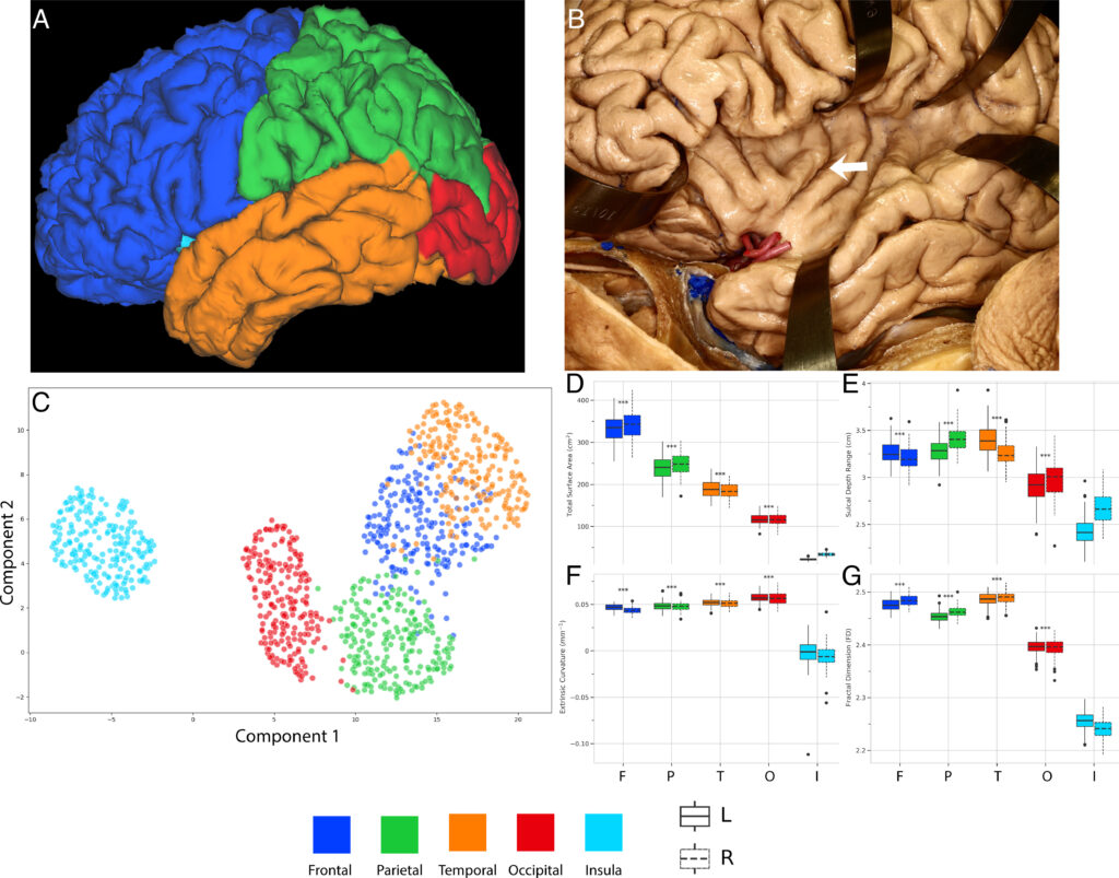
本研究は、島皮質が成体において他の葉とは異なる形態を持つことを定量的に立証し(図1)、これらの違いが胎児期の発達中に現れることを示した(図2)。具体的には、島皮質は他のどの脳葉よりも遅い速度で拡大し、接線方向拡大モデルで予測される幾何学的形状とは異なる、独特の回旋配置と形態を持つ。ヤコビアン分析により、体積成長の差は島皮質(低成長)とその周囲の眼球(高成長)の間で最も顕著であることが判明した(図2G)。この体積成長の顕著な差はシルヴィア裂を閉じ、前頭葉基底部と前頭葉前部を近接させ、大脳のC字形を確立する。接線方向の拡張の違いや、それに関連した皮質の拡張の制約といった既存の回旋モデルによって、接線方向の拡張の違い(例えば表面積の成長)が回旋パターンの違いに直接結びつけられている。その結果、島皮質における表面積(例えば接線方向)の拡大の欠如は、この葉の浅く比較的隆起していない表面と直接結びついている。
結論
島皮質における回形成と成長のユニークなパターンと、神経細胞の弯曲した移動軌跡を明らかにした。他のすべての終脳皮質とは対照的に、島皮質は、脳室体の小領域から移動し、レンズ状核の周りをカーブして島皮質に到達する、湾曲した放射状の移動流から形成される。この発達の違いが、島皮質の形態の変化と相対的に遅い成長の両方をもたらしたと考えられる。周囲の前頭葉、側頭葉、頭頂葉の過成長は、より大きな前駆領域から放射状にまっすぐ、妨げられることなく移動してくるため、島皮質の成長を上回り、シルビウス裂周辺の大脳の特徴的なC字型を形成する。
〇外側溝
外側溝は前頭葉、および頭頂葉と、側頭葉を上下に分けている。外側溝は大脳の両半球に存在するが、左半球のものの方が長い。外側溝はヒトの脳の発生の最も初期に形成される脳溝の1つであり、妊娠約14週目には見ることができる[1]。
外側溝は多くの側枝を持っている。最も顕著で多くの人で見つかるのは、外側溝上行枝 (垂直枝) と水平枝で、下前頭回を下位領域に分割する。また、外側溝は聴覚野の主要部位である横側頭回を含む。(Wkipedia)

“Journal Club (July 15, 2023)” への1件のフィードバック