Scientific Reports Published: 02 December 2014
Bipolar/rod-shaped microglia are proliferating microglia with distinct M1/M2 phenotypes (双極/棒状ミクログリアは、異なる M1/M2 表現型を持つ増殖性ミクログリアである)
Wing Yip Tam & Chi Him Eddie Ma
1Department of Biomedical Sciences, City University of Hong Kong, Tat Chee Avenue, Hong Kong (香港城市大学)
2Centre for Biosystems, Neuroscience, and Nanotechnology, City University of Hong Kong, Tat Chee Avenue, Hong Kong
3State Key Laboratory in Marine Pollution, City University of Hong Kong, Tat Chee Avenue, Hong Kong
香港城市大学:九龍塘に本部を置く香港の公立大学で1984年創立された。1994年大学設置。創設間もないが、すでにQSなどの世界大学ランキングでは上位に顔を出し始めているように、国際的に高く評価されている。(Wkipedia)
Abstract
Microglia are generally considered the resident immune cells in the central nervous system (CNS) that regulate the primary events of neuroinflammatory responses. Microglia also play key roles in repair and neurodegeneration of the CNS after injury. Recent studies showed that trains of bipolar/rod-shaped microglia align end-to-end along the CNS injury site during the initial recovery phase. However, the cellular characteristics of bipolar/rod-shaped microglia remain largely unknown. Here, we established a highly reproducible in vitro culture model system to enrich and characterize bipolar/rod-shaped microglia by simply generating multiple scratches on a poly-d-lysine/laminin-coated culture dish. Trains of bipolar/rod-shaped microglia formed and aligned along the scratches in a manner that morphologically resembled microglial trains observed in injured brain. These bipolar/rod-shaped microglia were highly proliferative and expressed various M1/M2 markers. Further analysis revealed that these bipolar/rod-shaped microglia quickly transformed into amoeboid microglia within 30 minutes of lipopolysaccharide treatment, leading to the upregulation of pro-inflammatory cytokine gene expression and the activation of Jak/Stat. In summary, our culture system provides a model to further characterize this highly dynamic cell type. We suggest that bipolar/rod-shaped microglia are crucial for repairing the damaged CNS and that the molecular mechanisms underlying their morphological changes may serve as therapeutic biomarkers.
ミクログリアは、一般に中枢神経系(CNS)に常駐する免疫細胞であり、神経炎症反応の主要な事象を制御していると考えられている。また、傷害後の中枢神経系の修復や神経変性においても、ミクログリアは重要な役割を担っている。最近の研究では、初期回復期において、双極性(bipolar)/棒状のミクログリアがCNSの損傷部位に沿って端から端まで整列することが示された。しかし、双極性/桿状ミクログリアの細胞特性はほとんど不明なままである。そこで、ポリリジン/ラミニンコートした培養皿に複数の傷をつけるだけで、双極性/桿状ミクログリアを濃縮・特性化できる再現性の高いin vitro培養モデル系を確立しました。その結果、傷ついた脳で観察されるミクログリアと同様の形態で、傷に沿った双極性/桿状ミクログリアの整列が形成された。これらの双極/棒状ミクログリアは、非常に増殖性が高く、様々なM1/M2マーカーを発現していました。さらに、これらの双極/桿状ミクログリアは、リポポリサッカライド処理後30分以内にアメーバ状ミクログリアに急速に変化し、炎症性サイトカイン遺伝子発現の上昇とJak/Statの活性化を引き起こすことが明らかになった。まとめると、我々の培養系は、この非常にダイナミックな細胞タイプの特徴をさらに解明するためのモデルを提供するものである。我々は、双極性/桿状ミクログリアが損傷した中枢神経系を修復するために重要であり、その形態的変化の根底にある分子メカニズムが治療バイオマーカーとして役立つ可能性があることを示唆している。
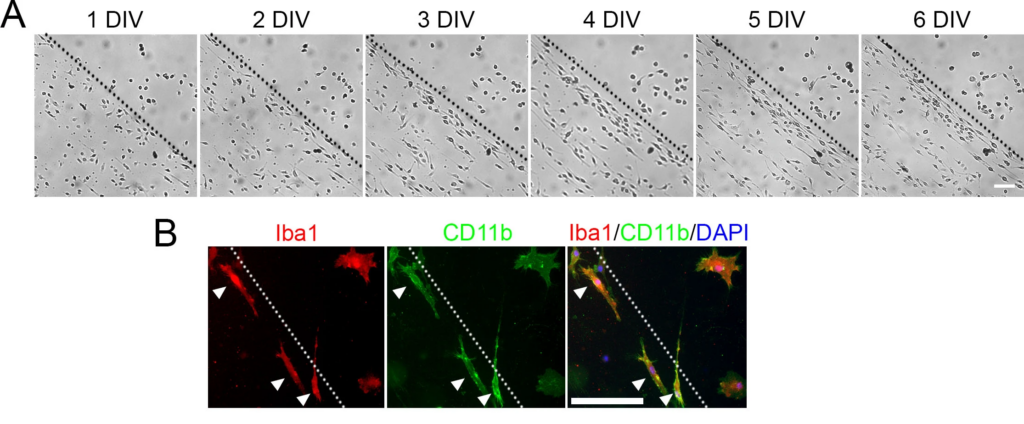
Bipolar/rod-shaped microglia aligned along the scratch of the coated surface in vitro.
(a) Bipolar/rod-shaped microglia colonized in the scratched area of a PDL/laminin-coated surface. The cell processes were randomly aligned at 1 DIV and then displayed a more synchronized alignment along the scratch at 6 DIV. (b) Both bipolar/rod-shaped (arrows) and amoeboid microglia expressed the microglial markers Iba1 (red) and CD11b (green). The dotted line indicates the boundary between the scratched (left to the line) and non-scratched areas. Scale bars: A: 100 μm; B: 50 μm.
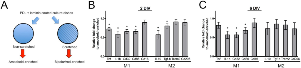
Reduced expression of M1 and M2 markers in bipolar/rod-shaped microglia.
(a) Schematic diagram illustrating the amoeboid-enriched and bipolar/rod-shaped enriched cultures. (b) Reduced expression of selected M1 and M2 markers in the bipolar/rod-enriched microglia cultures compared with the amoeboid-enriched cultures at 2 DIV (n = 5 to 6). (c) This reduced M1 marker expression was generally maintained, whereas M2 marker expression increased at 6 DIV (n = 5 to 6). *P < 0.05 based on Student’s t-test.
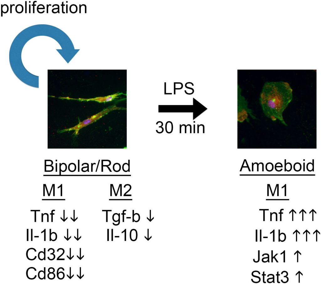
Schematic diagram illustrating the characterization of bipolar/rod-shaped microglia.
Bipolar/rod-shaped microglia are highly proliferative and expressed low levels of M1 and M2 markers. Upon LPS treatment, bipolar/rod-shaped microglia rapidly transformed into an amoeboid morphology and became M1-activated. (双極/桿状ミクログリアは増殖性が高く、M1およびM2マーカーを低レベルで発現している。LPS処理により、双極/桿状ミクログリアは急速にアメーバ状の形態に変化し、M1活性化された。)
Discussion
ミクログリアによる炎症性サイトカインの持続的な産生は、慢性的な神経炎症をもたらし、パーキンソン病やアルツハイマー病などの神経変性疾患の発症と関連している。我々のデータでは、双極/桿状ミクログリアは炎症性サイトカインであるTnfとIl-1bの発現量が少なく、双極/桿状ミクログリアは炎症による神経変性に寄与しないことが示唆された。しかし、双極/桿状ミクログリアにおける抗炎症性サイトカインIl-10とTgf-βの発現は、2DIVから6DIVにかけて増加した。IL-10はミクログリアによる炎症性サイトカイン産生を抑制することが知られている。以上のことから、双極/桿状ミクログリアは、脳損傷部位において、炎症性サイトカインよりも抗炎症性サイトカインを大量に産生することにより、神経保護作用を発揮している可能性が考えられる。炎症性サイトカインと抗炎症性サイトカインの発現のバランスは、脳損傷からの回復の程度や神経変性疾患の進行を決定する。
ミクログリアとマクロファージは、その起源が異なるためか、M1マーカーとM2マーカーの異なる発現プロフィールを示す。蓄積された証拠から、もともとマクロファージに採用されていたミクログリアのM1とM2への分類という概念は、ますます議論の的になっていることが示唆される6,7,12。研究により、いくつかのM1またはM2マーカーはマクロファージにのみ発現し、ミクログリアには発現しないことが明らかになっている。例えば、ヒトのM2活性化ミクログリアは、Arg1、Chi3l3、CD23、CD163、CD2067などのM2マクロファージマーカーを発現しなかった。生後脳の発達過程では、ミクログリアはM1とM2の両方のマーカーを発現しており、未熟なミクログリアはM1またはM2の表現型にコミットしないことが示唆されている。中枢神経系の炎症を引き起こす化学誘引タンパク質であるCCL2は、ミクログリアにおいて炎症反応と抗炎症反応の両方の発現を誘導し、それによってM1にもM2にも偏らない表現型を示している。この結果は、ナイーブな双極性/棒状ミクログリアがM1またはM2マーカーを独占的に発現するのではなく、LPSによる活性化の直後にM1表現型に容易に変化することを示した我々のデータとよく一致する。驚くべきことに、LPSによる形態変化と炎症性サイトカイン/Jak1/Stat3遺伝子発現のアップレギュレーションの両方が、約30分以内に検出された。したがって、双極性/桿状型ミクログリアは炎症性サイトカイン産生には関与しないが、LPS刺激により急速に活性型に変化し、高レベルの炎症性サイトカインを産生することが示唆された。
図6にまとめたように、PDL/ラミニンを塗布した培養皿に傷をつけると、M1でもM2でもない表現型にコミットし、高い増殖性を持つ双極性/ロッド型ミクログリアの形成が促進されることが示された。LPS処理により、双極/桿状ミクログリアは急速にアメーバ状に変化し、M1活性化することがわかった。このin vitro培養系は、双極性/桿状ミクログリアにおける遺伝子発現研究およびM1/M2マーカーのさらなる機能的特徴付けに利用でき、神経疾患に対する治療の可能性を示すことが期待される。
〇LPS LPSは、グラム陰性細菌の成分で、グラム陰性細菌の細胞壁の外側に埋め込まれた形で存在している。糖と脂質が結合した構造をしているので、「糖脂質」あるいは「リポ多糖」と呼ばれ、英語では「リポポリサッカライド(Lipopolysaccharide)」、略してLPSと呼ばれる。