Expansion sequencing: Spatially precise in situ transcriptomics in intact biological systems (拡大シーケンス:生物システム上の空間的に精確なin situトランスクリプトーム)
Shahar Alon, Daniel R. Goodwin, Adam H. Marblestone, Edward S. Boyden et al.
- Department of Media Arts and Sciences, MIT, Cambridge, MA, USA.
- McGovern Institute, MIT, Cambridge, MA, USA. マサチューセッツ工科大学マクガヴァン研究所
Science 29 Jan 2021: Vol. 371, Issue 6528, eaax2656. DOI: 10.1126/science.aax2656
*Scienceは、1880年に創刊された、アメリカ科学振興協会 (AAAS)によって発行されている学術雑誌である。
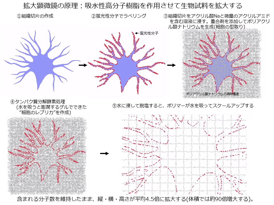
*単一細胞RNA-seqでは、組織から細胞や細胞核を解離する必要があるので、その細胞が存在していた解剖学的・空間的な位置情報が消去してしまう。in situ sequence法は、生物組織における複数のmRNAの分布を情報処理技術も併用して同定する方法である。現在のところ、大きい組織の空間トランスクリプトミクスの空間解像度は限定されている。本論文では、拡大顕微鏡で組織を膨潤させてからin situ sequencingすれば空間解像度の問題点を解決できるのではないか?という発想に基づくアプローチをとっている。
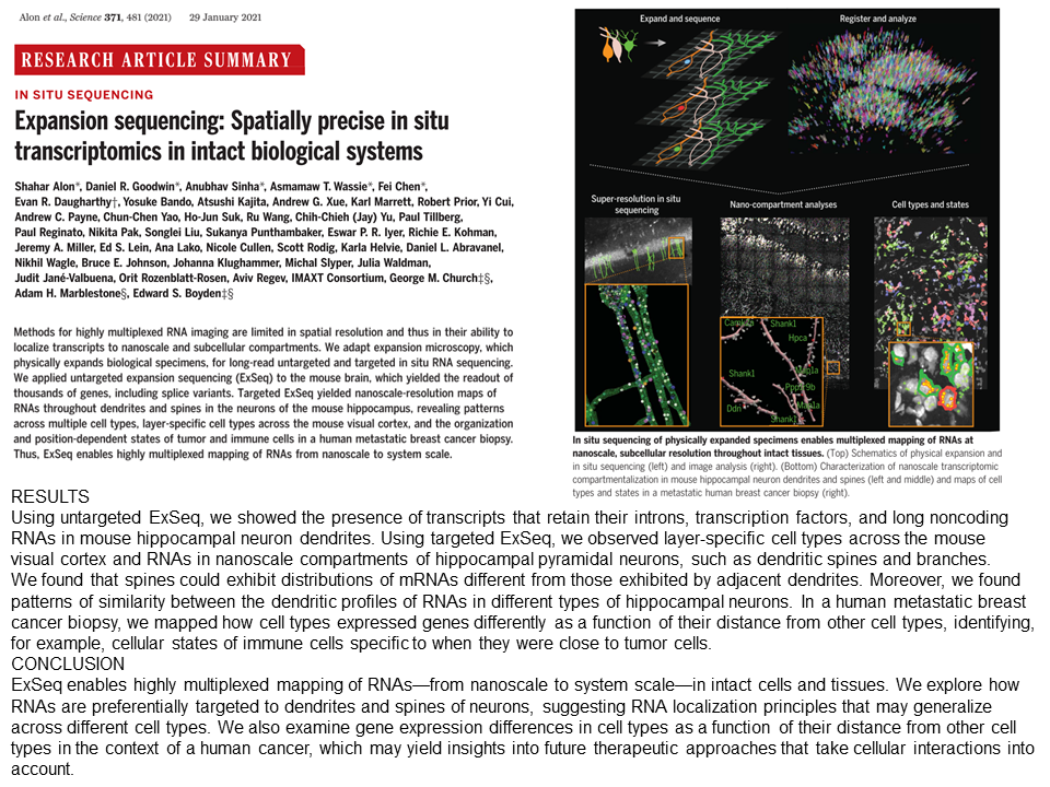
Identifying transcript location in cells
Identifying where specific RNAs occur within a cell or tissue has been limited by technology and imaging capabilities. Expansion microscopy has allowed for better visualization of small structures by expanding the tissues with a polymer- and hydrogel-based system. Alon et al. combined expansion microscopy with long-read in situ RNA sequencing, resulting in a more precise visualization of the location of specific transcripts. This method, termed “ExSeq” for expansion sequencing, was used to detect RNAs, both new transcripts and those previously demonstrated to localize to neuronal dendrites. Unlike other in situ sequencing methods, ExSeq does not target sets of genes. This technology thus unites spatial resolution, multiplexing, and an unbiased approach to reveal insights into RNA localization and its physiological roles in developing and active tissue.
細胞または組織内で特定のRNAがどこで発生しているかを特定することは、技術やイメージングによって制限されてきた。拡大顕微鏡法(Expansion Microscopy法)は、おむつなどにも使用されるポリマーおよびハイドロゲルベースのシステムであり、組織を膨張させることにより、小さな構造を可視化することを可能にしてきた。本論文で、マクガヴァン脳研究所(MIT)のBoydenとAlonらは、拡大顕微鏡法とlong-readのin situ RNAシーケンス法を組み合わせ、特定の転写物の位置をより正確に可視化することに成功した。この方法は、拡張シークエンスのための「ExSeq」と呼ばれるもので、新しい転写産物と以前にニューロンの樹状突起に局在することが証明された転写産物の両方のRNAを検出するために使用された。他のin situシーケンシング法とは異なり、ExSeqは遺伝子セットを標的としていない。この技術は、空間分解能、多重化、偏りのないアプローチを統合し、発生過程あるいは活動している組織におけるRNAの局在とそれらの生理的な役割を明らかにすることができる。
マクガヴァン脳研究所(MIT)のBoydenらによる拡大顕微鏡についてのオリジナル論文(Science. 2015)はこちら。
Expansion microscopy https://science.sciencemag.org/content/347/6221/543
拡大顕微鏡についての動画解説はこちら。