本日は、医療科学類の学生の方と一緒に以下の論文を勉強しました。
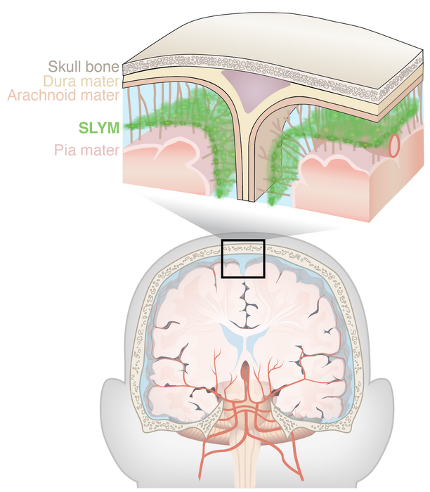
コペンハーゲン大学とロチェスター大学の研究グループは,クモ膜下腔に脳脊髄液(CSF)を2区画に分ける薄い膜が存在することを発見した。これまで髄膜は硬膜・クモ膜・軟膜の3層から形成されているとされてきたが、「第4の髄膜」の存在が示された。

Science. 2023 Jan 6;379(6627):84-88. doi: 10.1126/science.adc8810. Epub 2023 Jan 5.
A mesothelium divides the subarachnoid space into functional compartments (クモ膜下腔を機能的に分割する中膜)
Kjeld Møllgård # 1, Felix R M Beinlich # 2, Peter Kusk # 2, Leo M Miyakoshi # 2, Christine Delle 2, Virginia Plá 2, Natalie L Hauglund 2, Tina Esmail 2, Martin K Rasmussen 2, Ryszard S Gomolka 2, Yuki Mori 2, Maiken Nedergaard 3
- 1Department of Cellular and Molecular Medicine, Faculty of Health and Medical Sciences, University of Copenhagen, 2200 Copenhagen, Denmark.
- 2Division of Glial Disease and Therapeutics, Center for Translational Neuromedicine, Faculty of Health and Medical Sciences, University of Copenhagen, 2200 Copenhagen, Denmark.
- 3Division of Glial Disease and Therapeutics, Center for Translational Neuromedicine, University of Rochester Medical Center, Rochester, NY 14642, USA.
#Contributed equally.
An extra layer lines the brain
The traditional view is that the brain is surrounded by three layers, the dura, arachnoid, and pia mater. Møllgård et al. found a fourth meningeal layer called the subarachnoid lymphatic-like membrane (SLYM). SLYM is immunophenotypically distinct from the other meningeal layers in the human and mouse brain and represents a tight barrier for solutes of more than 3 kilodaltons, effectively subdividing the subarachnoid space into two different compartments. SLYM is the host for a large population of myeloid cells, the number of which increases in response to inflammation and aging, so this layer represents an innate immune niche ideally positioned to surveil the cerebrospinal fluid. —SMH
脳に並ぶ”余分な”髄膜層
従来、脳は硬膜、クモ膜、軟膜の3つの層で囲まれていると考えられていた。Møllgårdらは、クモ膜下リンパ様膜(SLYM: the subarachnoid lymphatic-like membrane)と呼ばれる第4の髄膜の層を発見した。SLYMは、ヒトおよびマウスの脳の他の髄膜層とは免疫表現型が異なっており、3kD以上の溶質に対する堅固な障壁を形成して、クモ膜下腔を実質的に2つの異なる区画に区分している。SLYMは骨髄系細胞の大きな集団の宿主であり、その数は炎症と老化に反応して増加するため、この層は脳脊髄液を監視するのに理想的な自然免疫ニッチであるといえる。
Abstract
The central nervous system is lined by meninges, classically known as dura, arachnoid, and pia mater. We show the existence of a fourth meningeal layer that compartmentalizes the subarachnoid space in the mouse and human brain, designated the subarachnoid lymphatic-like membrane (SLYM). SLYM is morpho- and immunophenotypically similar to the mesothelial membrane lining of peripheral organs and body cavities, and it encases blood vessels and harbors immune cells. Functionally, the close apposition of SLYM with the endothelial lining of the meningeal venous sinus permits direct exchange of small solutes between cerebrospinal fluid and venous blood, thus representing the mouse equivalent of the arachnoid granulations. The functional characterization of SLYM provides fundamental insights into brain immune barriers and fluid transport.
中枢神経系は、古典的には硬膜、クモ膜、軟膜と呼ばれる髄膜で覆われている。我々は、マウスとヒトの脳において、くも膜下空間を区画する第四の髄膜層が存在することを示し、クモ膜下リンパ様膜(SLYM: SLYM: the subarachnoid lymphatic-like membrane)と名づけた。SLYMは、形態学的および免疫表現学的に、末梢臓器や体腔を覆う中皮膜に類似しており、血管を包み込み、免疫細胞を保持している。機能的には、SLYMは硬膜静脈洞の内皮に密着しているため、脳脊髄液と静脈血の間で小さな溶質の直接交換が可能であり、マウスにおけるクモ膜顆粒(ヒトでは観察される)に相当するものである。SLYMの機能解析は、脳の免疫バリアと体液輸送に関する基本的な知見を提供するものである。
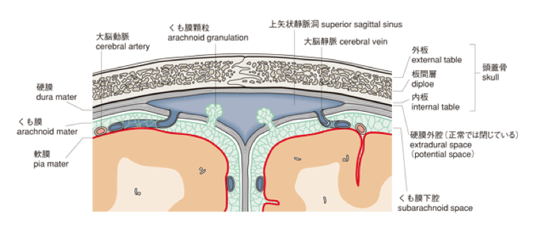
*クモ膜顆粒(arachnoid granulation; パキオニ小体)とは静脈洞に突出する中皮の塊をさす。脳のクモ膜のある部分ではその外面が硬膜に向かって桑の実のような膨隆をつくり、硬膜と融合して硬膜静脈洞に突出しクモ膜顆粒を形成している。くも膜顆粒はくも膜の外面に並ぶ中皮が集まり、結節状に盛り上がったもので中皮由来の細胞が単層または重層になって果粒の表面をおおい、内部には結合組織のふくらみがみられる。(Wkipedia)
Introduction
脳脊髄液(CSF)が、中枢神経系において準リンパ系として働くという概念を支持する新たな証拠が得られている。心血管系の拍動性により、髄液は動脈周囲腔に沿って脳深部へと流入し、そこでグリアアクアポリン4(AQP4)水チャネルによって促進される髄液と間質液との交換が行われる。ニューロピルからの体液と溶質は、静脈周囲腔や脳神経を含む複数の経路に沿って除去され、最終的には髄膜および頸部リンパ管を介して静脈循環に輸送される。髄液の再吸収は、くも膜顆粒を介して静脈洞でも起こる可能性があるが、げっ歯類ではまだ報告されていない。リンパ管-リンパ管経路に沿った髄液の流れを研究する努力にもかかわらず、クモ膜下腔という大きな空洞の中で髄液がどのように輸送されるかはまだ明らかにされていない。本研究では、マウスとヒトの脳を取り囲むクモ膜下腔内で、髄液と免疫細胞の輸送がどのように組織化されているかを調べた。
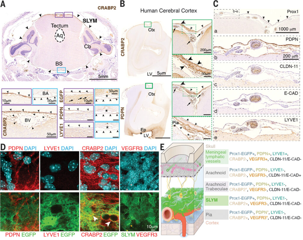
(A) Sections of Prox1-EGFP+ mouse brain after decalcification of the whole head counterstained with Mayer’s hematoxylin (M-HE, purple) show that SLYM (arrowheads) is positively immunolabeled for CRABP2 (brown) and Prox1-EGFP+/PDPN+/LYVE1−/VEGFR3− and encases the entire brain, covering its dorsal and ventral portions (purple and blue insets, respectively). (B) Adult human brain sections immunolabeled for CRABP2 and PDPN reveal the presence of SLYM (arrowheads) that enwraps the subarachnoid space blood vessels (arrow). Ependymal and pia mater cells are also PDPN+ (asterisks). (C) Serial sections of the same adult human material immunolabeled for Prox1, PDPN, CLDN11, E-CAD, and LYVE1. SLYM is indicated by arrowheads. (D) Confocal images of SLYM immunolabeling showing positive labeling for PDPN and CRABP2 (both in red). No signal was detected for LYVE1 or VEGFR3. (E) Schematic representations of the immunophenotypical characterization of the meningeal layers, meningeal lymphatic vessels, and arachnoid trabeculations. For arachnoid trabeculae, CRABP2* signifies that the trabeculae are CRABP2− in the outer SAS but CRABP2+ in the inner SAS. For pia, PDPN* indicates that pia is PDPN+ in many regions of pia, but not all. VEGFR3* signifies that pia was VEGFR3+ only in a few regions. Aq, aqueduct; BA, basilar artery; BS, brain stem; BV, blood vessel; Cb, cerebellum; Ctx, cerebral cortex; Ep, ependyma; LV, lateral ventricle.
https://www.science.org/doi/10.1126/science.adc8810?url_ver=Z39.88-2003&rfr_id=ori:rid:crossref.org&rfr_dat=cr_pub%20%200pubmed
Discussion
脳を覆う髄膜の重要な役割が認識されるようになったのは最近のことである。髄膜のリンパ管網によって髄液が排出され、この排出が抑制されると、神経変性の動物モデルにおいてタンパク質の凝集と認知機能の低下が促進されることが知られている。SLYM(スリム)はProx1+/PDPN+/LYVE1-/CRABP2+/VEGFR3-/CLDN-11-/E-Cad-であり、硬膜、くも膜、梨状膜などの従来の髄膜、髄膜リンパ管、くも膜海綿体とは異なる。SLYMはくも膜下腔を2つの区画に細分化しており、髄液の輸送が現在考えられているよりも組織化されていることを示唆している。例えば、内側のくも膜下腔の血管系を覆っているSLYMは、外側のくも膜下腔コンパートメントに存在する溶質を循環させることなく、貫通する細動脈に沿って脳実質へとCSF流入を導く。しかし、第4の髄膜層であるSLYMが発見されたことは、体液輸送以外にもいくつかの意味を持つ。SLYMが分子量3kDa以上の髄液性溶質のバリアであるという観察は、より詳細な研究を必要とするが、SLYMを含めてCNSバリアの概念を再定義する必要性を示している。髄膜はCNSの免疫監視を担う骨髄系細胞の宿主であり、SLYMは脳表面と密接に関連していることから、この監視において重要な役割を果たしていると考えられる。ここで我々は、急性炎症と自然老化に応答して、SLYMに存在する免疫細胞の数と多様性が大きく増加することを示した。SLYMの物理的な破裂は、髄液流動パターンを変化させることにより、外傷性脳損傷後のリンパ液流動の長期的な抑制や、アルツハイマー病発症の外傷後リスクの上昇を説明できる可能性がある。また、SLYMが破裂すると、頭蓋骨の骨髄からくも膜下腔内部へ免疫細胞が直接通過し、脳表面へ直接アクセスできるようになるため、外傷性脳損傷後の神経炎症が長引くことが説明できる可能性がある。SLYMは、多くの免疫細胞の宿主であるだけでなく、CNS免疫にも直接関与している可能性がある。リンパ様組織は炎症が起きると急速に変化するが、これは脳において、多発性硬化症などの疾患と顕著な関連性を持つ可能性がある。
20200522-Presentation-01ネダーガード教授は,2012年に発表した脳内老廃物の排出機構「グリンパティックシステム」の発見者である。「脳のリンパ系」とも呼ばれるこのシステムは、睡眠時にCSFの流入を促して「脳の清掃」を加速させ、またこのシステムがダメージを受けると脳損傷後の治癒が阻害され、アルツハイマー病など神経変性疾患の原因となる毒性タンパク質の蓄積につながると考えられている。
Summary
SLYMはクモ膜下腔を2つの区画に区分し,分子の交換を制限している。このことからCSF循環はこれまで考えられてきたよりも複雑に組織化されていることが示唆される。SLYMは以下の特徴をもつ。
・クモ膜下腔を二分する、厚さ14 μm程度の単層の中皮膜である。
・他の髄膜とは免疫表現型が異なる特徴を持ち、脳脊髄液が満たされる2つの区画を機能的に隔てる。
・頭部運動時などの機械的ストレスから脳を守る。
・CSF・グリンパティック系の排出ルートとしての可能性がある。
・免疫細胞をSLYM内に動員し、脳を監視・防御する最前線の役目を持っている。
睡眠中には脳内から“毒素”が洗い流される:米研究チームがメカニズムを解明、アルツハイマー病の治療に光

“Journal Club (January 23, 2023)” への1件のフィードバック