Nature Communications volume 12, Article number: 5289 (2021)
Open Access
Published: 06 September 2021
Capillary-associated microglia regulate vascular structure and function through PANX1-P2RY12 coupling in mice (毛細血管関連ミクログリアはPANX1-P2RY12結合を介して血管の構造と機能を制御する)
Kanchan Bisht, Kenneth A. Okojie, Kaushik Sharma, Dennis H. Lentferink, Yu-Yo Sun, Hong-Ru Chen, Joseph O. Uweru, Saipranusha Amancherla, Zainab Calcuttawala, Antony Brayan Campos-Salazar, Bruce Corliss, Lara Jabbour, Jordan Benderoth, Bria Friestad, William A. Mills III, Brant E. Isakson, Marie-Ève Tremblay, Chia-Yi Kuan & Ukpong B. Eyo
*バージニア大学は、1819年に創設されたアメリカ合衆国バージニア州シャーロッツビルに本部を置くアメリカの州立大学である。
Abstract
Microglia are brain-resident immune cells with a repertoire of functions in the brain. However, the extent of their interactions with the vasculature and potential regulation of vascular physiology has been insufficiently explored. Here, we document interactions between ramified CX3CR1 + myeloid cell somata and brain capillaries. We confirm that these cells are bona fide microglia by molecular, morphological and ultrastructural approaches. Then, we give a detailed spatio-temporal characterization of these capillary-associated microglia (CAMs) comparing them with parenchymal microglia (PCMs) in their morphological activities including during microglial depletion and repopulation. Molecularly, we identify P2RY12 receptors as a regulator of CAM interactions under the control of released purines from pannexin 1 (PANX1) channels. Furthermore, microglial elimination triggered capillary dilation, blood flow increase, and impaired vasodilation that were recapitulated in P2RY12−/− and PANX1−/− mice suggesting purines released through PANX1 channels play important roles in activating microglial P2RY12 receptors to regulate neurovascular structure and function.
ミクログリアは、脳に常駐する免疫細胞であり、脳内で様々な機能を発揮している。しかし、ミクログリアが血管系とどのように相互作用し、血管の生理機能を制御しているのかについては十分に検討されていない。ここでは、ラミファイド型のCX3CR1+骨髄系細胞体と脳毛細血管との相互作用を記録した。これらの細胞が正真正銘のミクログリアであることを、分子的、形態学的、超微細構造学的アプローチによって確認した。次に、これらの毛細血管関連ミクログリア(CAM)の詳細な時空間的特徴を、ミクログリアの削除時および再増殖時を含む形態学的活動において実質ミクログリア(PCM)と比較して示す。分子的には、P2RY12受容体が、パンネキシン1(PANX1)チャネルから放出されるプリンを制御することで、CAMの相互作用を調節していることを明らかにした。さらに、ミクログリアの除去によって引き起こされる毛細血管の拡張、血流の増加、血管拡張の障害は、P2RY12-/-マウスとPANX1-/-マウスで再現された。このことから、PANX1チャネルから放出されるプリンがミクログリアのP2RY12受容体を活性化し、神経血管の構造と機能を制御する上で重要な役割を果たしていると考えられる。
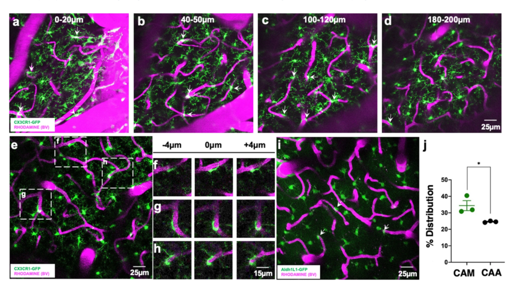
a–d Representative 20-µm-thick two-photon projection images from a CX3CR1GFP/+ adult brain showing myeloid cells (green) and the vasculature (rhodamine in magenta) at varying tissue depths between the brain surface and 200 µm of the cortex. Arrows identify capillary-associated ramified myeloid cells. e Representative 20-µm-thick in vivo two-photon image from a CX3CR1GFP/+ adult brain showing ramified myeloid cells (green) and the vasculature (rhodamine in magenta) in the cortex. f–h Representative images from boxed regions in (e) showing somal interactions between the ramified myeloid cells and capillaries in an 8 µm tissue volume. i, j Representative 20-µm-thick two-photon projection image (i) and quantification (j) from an ALDH1L1GFP/+ P30 brain showing astrocytes (green) and the vasculature (rhodamine in magenta) in the cortex. Capillary-associated astrocytes (CAAs, arrows in i) density is compared to capillary-associated myeloid (CAM) density. n = 3 mice each. Representative images in (a–h) were observed in five mice and in (i) was observed in three mice. Data are presented as mean values ± SEM. *p < 0.05. Two-sided unpaired Student’s t test.
“Journal Club (September 27, 2021)” への1件のフィードバック